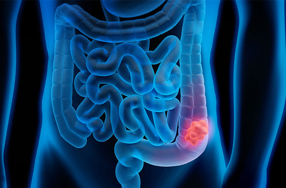
What is Rectum Cancer?
It is known that there are different "risk factors" that are effective in the development of colorectal cancer (CRC). These; being over 50 years old – due to fat
rich, fiber-poor diet – Presence of “synchronous” or “metachronous” (developed later) colorectal adenoma or CRC – “Amsterdam-I” “Amsterdam-II”, which relates cancer cases in the family to the possible cancer risk of other family members ” and “Hereditary risk factors” expressed by “Bethesta criteria” – can be counted as the “risk of developing CRC” that develops in the advanced stages of ulcerative colitis and Crohn's disease.
Gelişmiş toplumlarda 100 000’de 50 sıklık ile görülen ve tüm kanserler arasında 4. sırada yer alan KRK’lerin, yaklaşık %20’sini Rektum kanseri (RK) oluşturur. Rektumu kolon’dan ayıran ana özellik, yerleşiminin farklılığıdır.
RK leri, makroskopik patoloji açısından bakıldığında çoğunlukla “polipoid (vejetan) – ülseröz – infiltratif” tiplerde görürüz, en az rastlanan infiltratif tiplerdir. Mikroskopik olarak ise %98’i “adenokanser”dir.
Symptoms
While some patients with RC may remain asymptomatic until the disease reaches an advanced stage, they may consult a physician with complaints such as changes in defecation patterns (newly occurring constipation or diarrhea, recurrent), rectal bleeding, decrease in stool diameter, difficulty in defecation, and similar complaints. Perianal pain - anal sphincter, nerve tissues or bone involvement, and tenesmus (painful feeling of defecation) are symptoms of "advanced disease", suggesting a decrease in rectal capacity caused by a large-volume tumor. Stenosis that prevents passage through this area causes abdominal swelling, nausea, vomiting and colic-like pain. In some cases, the rectum may become obstructed by a tumor mass and a "bowel obstruction" situation may develop.
The most common symptom due to RC is change in defecation habits, and the second most common symptom is rectal bleeding.
Tumors can be detected by examining the anal canal and rectum with "finger" or "endoscopy".
Definitive Diagnosis
It is diagnosed by “endoscopy” and “biopsy”.
Carcinoembryonic antigen (CEA) is measured before surgery and used for comparison in postoperative follow-ups.
En önemli prognostik gösterge, teşhis esnasında “tümörün yaygınlık” derecesidir. İn situ karsinom (Tis), “yüksek grade displazi” olarak da kabul edilmektedir ve lenf nodu invazyonu görülmez. Barsak duvarına sınırlı (T1, T2) tümörlerde lenf nodu metastazı sırasıyla %5 ve %20 oranında görülürken rektum duvarını tamamen kaplayan ve komşu yapılara da taşan tümörlerde (T3, T4) lenf nodu tutulumu %50’yi aşmaktadır. Tutulan lenf nodu sayısı 4’ü aştığında, prognoz olumsuz etkilenir. KRK’de en sık rastlanan uzak metastaz karaciğerde görülür ve portal ven dallarının invazyonu yoluyla hematojen yayılıma bağlıdır.